
Macular Degeneration (Age-Related Macular Degeneration)
Known in medical literature as "Macular Degeneration," yellow spot disease is a condition where the macula (yellow spot) region, located at the back of the eye and responsible for sharp, clear, and detailed vision, becomes damaged over time.

Diabetic retinopathy is a condition where the capillaries of the retina—the inner layer at the back of the eye that transmits visual signals to the brain—are damaged due to blood sugar remaining high and uncontrolled for a long time.

The retina (nerve layer), which covers the back inner wall of our eye like wallpaper, consists of millions of nerve cells that perceive light from the outside and transmit it to the brain via optic nerves.
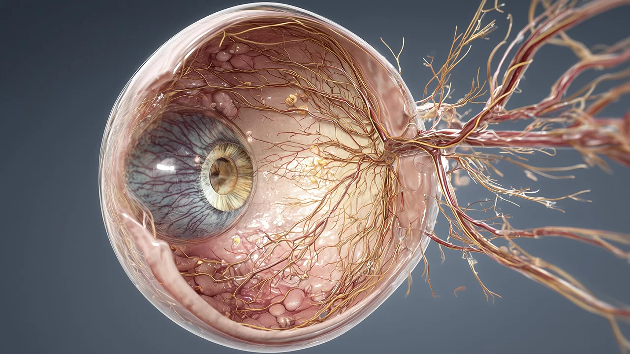
Just as our heart or brain needs an uninterrupted blood flow to function, the nerve layer of our eye (retina) similarly requires rich oxygen and nutrient support. The retina is nourished by a main artery (artery) coming from the center and sends back used, deoxygenated blood through a main vein (vein) also exiting from the center.

Retinopathy of Prematurity (ROP)
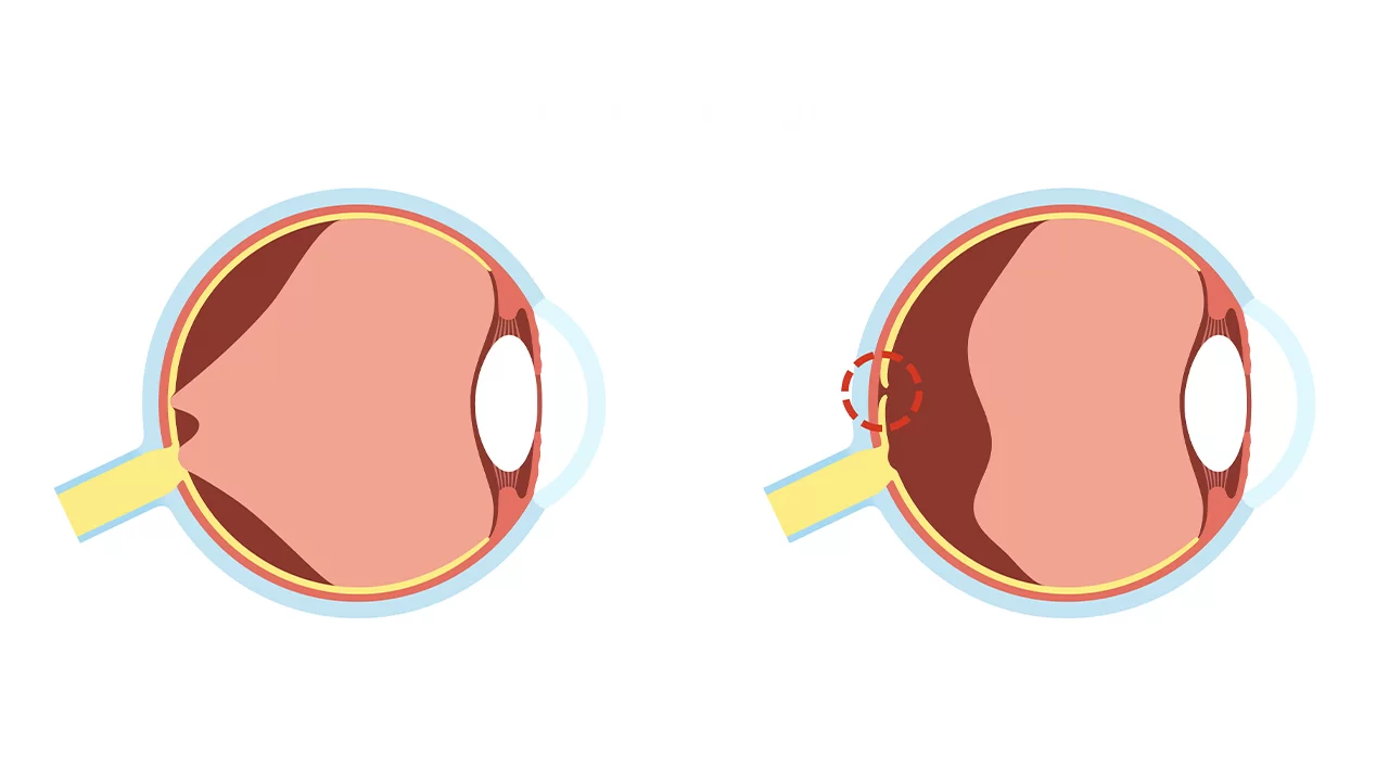
In a normal pregnancy process, vascular development in the baby's eye begins in the womb at approximately the 16th week and continues until birth (around the 40th week), reaching the far edges of the retina (the nerve layer of the eye). However, when a baby is born prematurely, this normal vascular development process is interrupted.
The nerve layer covering the back inner wall of our eye, consisting of light-sensitive nerve cells, is called the retina. The millimetric area located at the very center of the retina, approximately the size of a pinhead, is called the macula (yellow spot).

Also known in medical literature as "Macular Pucker," "Cellophane Maculopathy," or "Premacular Fibroplasia," Epiretinal Membrane (ERM) is a thin, semi-transparent, and membrane-like scar tissue that forms directly over the retina—the innermost layer of the eye—and specifically the macula (yellow spot) region responsible for sharp, detailed vision.

Central Serous Retinopathy (CSR)
The nerve layer covering the inner surface of our eye and initiating the vision process is called the retina. The most critical region of the retina is the macula (yellow spot), our sharp vision center responsible for functions such as reading, recognizing faces, and clearly distinguishing colors.

Retinitis Pigmentosa (RP) is not a single disease but a general name for a broad group of hereditary retinal dystrophies that cause the light-sensitive cells of the retina to degenerate slowly and irreversibly.

Known in medical literature as "Juvenile Macular Degeneration" or "Fundus Flavimaculatus," Stargardt Disease is a hereditary condition that directly targets the "macula" (yellow spot)—the center of the retina responsible for sharp, clear vision.

Leber Congenital Amaurosis (LCA)
First described in 1869 by Dr. Theodor Leber, Leber Congenital Amaurosis (LCA) is one of the most common genetic causes of congenital blindness in childhood. It is a group of severe and early-onset hereditary retinal dystrophies that cause the retina (the light-sensitive nerve layer at the back of the eye) to fail in its function from birth.

Congenital Retinal Dystrophies
In medicine, "congenital" means "present at birth," and "dystrophy" refers to a tissue or organ (in this case, the retina) failing to complete its normal development, becoming structurally impaired, and losing function due to genetic errors.

The nerve layer covering the back inner wall of our eye is called the retina, and the millimetric area located at the exact center of this layer, responsible for sharp, colored, and detailed vision, is called the "macula" (yellow spot). Reading books, driving, threading a needle, or recognizing a person's face depends entirely on the healthy functioning of the macula.

To maintain the vitality and spherical shape of the eyeball and to nourish avascular tissues like the cornea and lens, a special fluid called "aqueous humor" is constantly produced inside the eye. In a healthy eye, this fluid leaves through microscopic channels called the "trabecular meshwork" in the anterior chamber and enters the bloodstream.

Toxic Optic Neuropathies (Methyl Alcohol Intoxication)
Toxic Optic Neuropathy occurs when poisonous chemicals, heavy metals, or certain powerful drugs reach the eye via the bloodstream, poisoning and killing these vital nerve fibers at a cellular level. The most severe, rapid, and unfortunately, most lethal subtype of this condition is Methyl Alcohol (Methanol) Intoxication.

The optic nerve is a massive network of approximately 1.2 million fine nerve fibers that collects light and color signals formed in the retina (the back layer of the eye) and transmits them to the visual center (occipital cortex) at the back of the brain.